Pairs well with20 on file
Why these belong in the same formula. Each row says what the basis is, from settled biochemistry through to a trial that measured the pair.
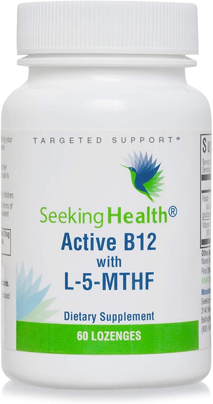
Folate and B12 run the same reaction: the enzyme methionine synthase uses B12 to pass folate's methyl group to homocysteine, which rebuilds methionine and frees folate to keep cycling. Without enough B12 the methyl group stays locked on folate and the cycle stalls, which is why the two are routinely supplied together.
Folate and B12 recycle homocysteine back into methionine, while vitamin B6 clears it down the other branch as the cofactor that carries homocysteine toward cysteine. Supplying all three supports the body's normal handling of homocysteine from both routes at once.
Riboflavin, in its active FAD form, is the cofactor for MTHFR, the enzyme that makes the 5-methyl form of folate the body uses to remethylate homocysteine. When riboflavin runs low, folate is converted into that active form less readily, so the two feed the same methylation step.
Betaine drives a second, folate-independent route for the same reaction: the enzyme BHMT uses betaine to remethylate homocysteine into methionine when the folate and B12 route is stretched. The two pathways back each other up within the body's normal methylation cycle.
Serine hydroxymethyltransferase moves a carbon from serine onto tetrahydrofolate, which is how most one-carbon units enter the folate cycle. Without serine there is little for folate to carry.
The glycine cleavage system loads a carbon onto tetrahydrofolate, and glycine N-methyltransferase pulls methyl groups back off the cycle. Glycine sits on both ends of the traffic folate handles.
Choline is oxidised to betaine, which can remethylate homocysteine without folate involvement. When folate intake is low, demand shifts onto choline, and the same is true in the other direction.
Betaine homocysteine methyltransferase uses trimethylglycine to remethylate homocysteine, running beside the folate and B12 route. Supplying both means the cell has either path available.
Folate has to be reduced and then methylated by MTHFR to become 5-methyltetrahydrofolate, the form found in plasma. Methylfolate is that end product supplied directly.
5-methyltetrahydrofolate hands its methyl group to homocysteine to make methionine, which is then activated to SAM. SAM levels also feed back on the folate cycle by regulating MTHFR.
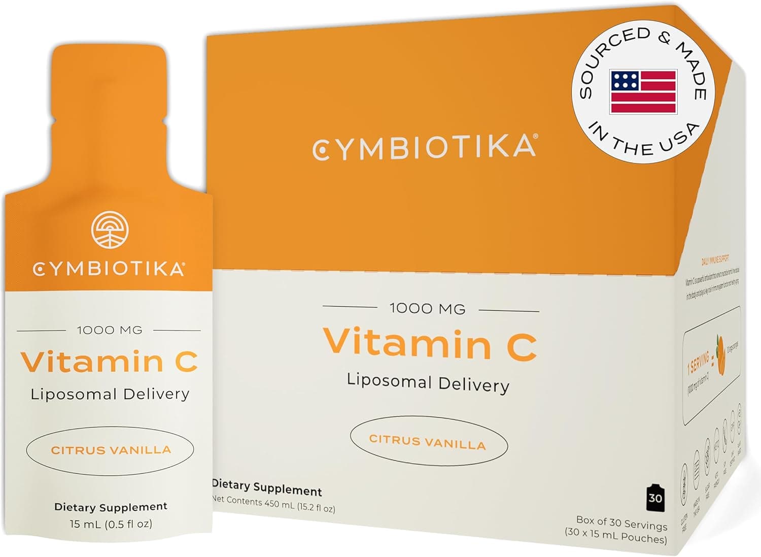
Reduced folates oxidise readily in the gut and in plasma, which inactivates them. Ascorbate maintains the reducing conditions that keep more of the dose intact.
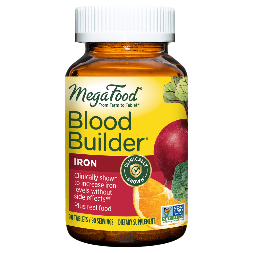
Folate supplies the one-carbon units for DNA synthesis in dividing marrow cells while iron is built into haem. A shortfall in either limits normal red cell production, which is why formulations pair them.
Intestinal folate conjugase is a zinc-dependent enzyme, so poor zinc status limits how much dietary polyglutamate folate can be absorbed. In the other direction, large folate doses can form complexes with zinc in the lumen and reduce its uptake.
Folate + L-MethionineEstablished one-carbon biochemistry: 5-methyltetrahydrofolate donates its methyl group to homocysteine to regenerate methionine, so folate status and methionine load sit on the same cycle. Methionine synthase pulls the methyl group off 5-methyltetrahydrofolate and puts it onto homocysteine, which reforms methionine. A larger methionine intake raises the amount of homocysteine cycling through that step, and folate supply is what allows the remethylation half to keep up. This is settled biochemistry rather than a combination trial, so it describes a pathway relationship and not a measured joint effect.
Folate + L-HistidineHistidine catabolism routes formiminoglutamate through tetrahydrofolate, the basis of the classic urinary FIGLU response to low folate status. Breaking histidine down to glutamate requires tetrahydrofolate to accept a formimino group. When folate supply is short, the intermediate accumulates and spills into urine, which is why histidine loading was used historically as a functional read on folate status. The relationship is textbook enzymology. It is not a claim that the two together produce an added benefit.
Folate + Vitamin B3 (niacin)NADPH, made from niacin-derived NADP, is the reducing cofactor for dihydrofolate reductase and for methylenetetrahydrofolate reductase. Folic acid has to be reduced twice before it enters the active folate pool, and both reductions spend NADPH. Methylenetetrahydrofolate reductase also runs on NADPH alongside its flavin cofactor. Niacin therefore sits upstream of the reducing power folate handling needs, which is a cofactor relationship rather than an outcome measured in a joint trial.
Folate + L-CysteineHomocysteine generated in the folate-dependent methylation cycle is disposed of through transsulfuration to cystathionine and then cysteine. Folate feeds the remethylation branch that turns homocysteine back into methionine. The other exit from homocysteine is transsulfuration toward cysteine, which needs vitamin B6. The two branches share one substrate pool, so cysteine sits downstream of the same junction folate acts on. Homocysteine is a blood marker of that junction, not a clinical outcome in itself.
Folate + Green tea extract (EGCG)Catechins including EGCG inhibit dihydrofolate reductase in laboratory systems and have been described as interfering with folate handling. EGCG binds dihydrofolate reductase in cell-free and cell-culture work, the same enzyme that converts folic acid into usable folate. Concentrated catechin extracts taken alongside folate are therefore worth spacing apart in the day. The evidence here is mechanistic and laboratory-based, not a human interaction trial.
Charcoal adsorbs a wide range of small organic molecules, and folate is a small water-soluble molecule that shares the gut lumen with it. Taking the two in the same window can reduce how much folate stays available for uptake, so separating doses is the usual practice. This is a general adsorption property rather than a folate-specific measurement.
Folate + Vitamin B6Established one-carbon biochemistry. Serine hydroxymethyltransferase is a PLP-dependent enzyme, and folic acid is among the highest co-study terms. Serine hydroxymethyltransferase, which loads a one-carbon unit onto tetrahydrofolate from serine, is PLP-dependent. B6 status therefore governs how well the folate cycle is supplied with one-carbon units. The two are inseparable in practice, which is why B-complex products carry both.